
Fragment-Based Drug Discovery 2.0, Part 1: Faster, Cleaner Weak-Binding Hit Discovery
By Mahmoud Khatib Al-Ruweidi
Fragment-based drug discovery (FBDD) emerged from the recognition that chemical space grows exponentially with molecular weight, making comprehensive screening of drug-sized molecules impossible.1 The rule of three formalized this insight: fragments with molecular weight below 300 Da, cLogP ≤3, and no more than three hydrogen bond donors, acceptors, or rotatable bonds sample chemical space efficiently while maintaining ligand efficiency.2 The strategy exploits an empirical design reality: fragments can start with high ligand efficiency (LE; ΔG_bind normalized by heavy-atom count), leaving headroom for potency growth as molecular weight increases — often at the cost of some efficiency erosion.3 In practice, a micromolar fragment with strong LE can be a better optimization substrate than a larger hit with weaker efficiency.3
Lead optimization benefits economically from fragment hits, which offer multiple ways to develop structure-activity relationships (SAR) simultaneously. Abbott Laboratories showed this by linking two weak-binding fragments, resulting in a 19 nM FKBP inhibitor with 5,000 times greater potency.4 This SAR by nuclear magnetic resonance (NMR) methodology established the paradigm: NMR detects binding, localizes sites, and enables rational design.
The Artifact Ecology: False Positives And False Negatives
The sensitivity required for fragment detection creates vulnerability to artifacts. High-throughput screening campaigns routinely encounter false-positive rates exceeding 90% from compound aggregation alone.5 Colloidal aggregates (50 to 500 nm) form above critical concentrations and nonspecifically adsorb enzymes, causing apparent inhibition. The pan-assay interference compounds (PAINS) framework highlights other interference sources, including redox cycling, metal chelation, covalent reactivity, and assay-specific fluorescence artifacts found in about 400 substructure classes.6
NMR reduces artifact issues by directly detecting binding, not relying on indirect measures like enzymatic turnover or fluorescence. It identifies binding-consistent line-shape behavior (chemical-shift changes and exchange broadening) and flags common artifact modes (e.g., aggregation/non-specific association) through nonlinear concentration dependence, unusually large/featureless linewidths, and poor reproducibility across orthogonal conditions, making artifacts recognizable to experts and clarifying results beyond what biochemical assays provide.
What "FBDD 2.0" Means: Three Technological Convergences
Three developments define the current era. First, engineered ¹⁹F fragment libraries exploit fluorine's unique NMR properties: 100% natural abundance, approximately 83% of ¹H's intrinsic sensitivity, negligible endogenous ¹⁹F background in most biological samples, and a chemical-shift range exceeding 200 ppm, enabling mixture screening of 20 to 36 compounds with manageable spectral overlap under library-specific mixture design.7
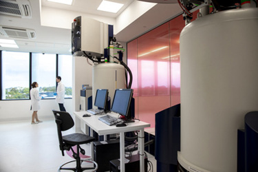
Second, ultra-high-field instruments have expanded from early adopters to operational deployments and new acquisitions: The Ohio State University installed the first U.S. 1.2 GHz spectrometer in December 2023,8 and Bruker announced in December 2024 that the Swiss High-field NMR Facility at the University of Zürich acquired a 1.2 GHz system.9 The 28.2 Tesla field strength provides resolution gains enabling larger fragment cocktails and sensitivity improvements benefiting protein-limited targets.
Third, contrast-engineered experiments improve ligand detection by optimizing relevant physics rather than just boosting signal strength. The PEARLScreen technique, developed at ETH Zürich, uses perfect-echo pulse trains with adjusted inter-pulse delays to detect binding through exchange-induced transverse relaxation, unlike conventional T₁ρ spin-lock methods.10 On the same screening setup, the required protein concentration could be reduced by up to one order of magnitude relative to conventional ligand-observed experiments; at 1.2 GHz, a 16-compound-mixture benchmark reached 40-fold lower protein consumption than a 600 MHz T₁ρ reference condition. These gains are benchmark-specific and depend on target size, exchange regime, mixture composition, and spectral resolution.
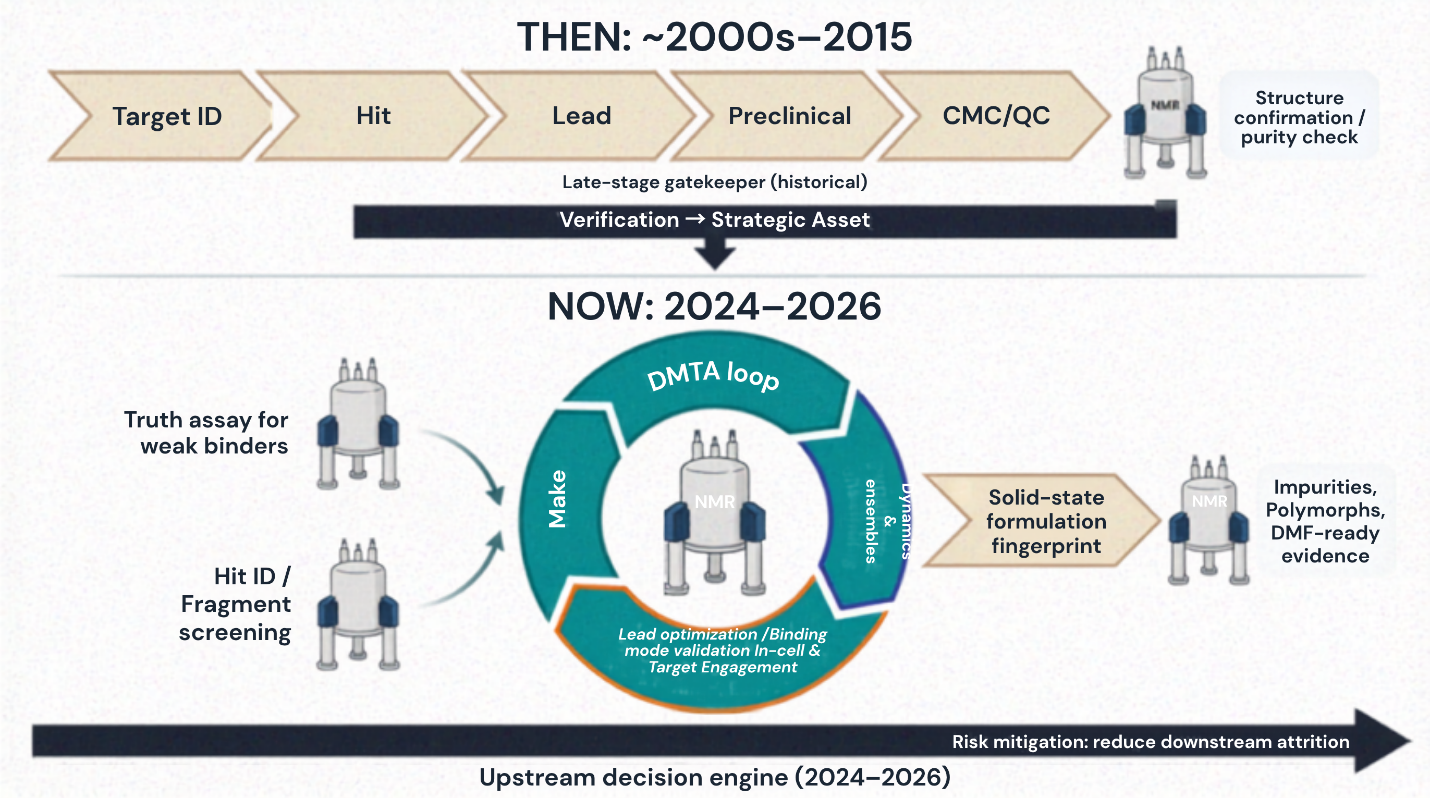
Figure 1. A summary of how NMR has shifted from a late-stage verification tool to an upstream decision engine integrated into Design-Make-Test-Analyze loops.
¹⁹F NMR For Discovery Teams: Three Modalities
Direct ¹⁹F cocktail screening
Most biological samples have negligible endogenous ¹⁹F background, so added fluorinated fragments can be detected with minimal spectral interference. Commercial ¹⁹F fragment libraries now include large, mixture-compatible sets with characterized chemical shifts, simplifying spectral assignment. Positioning fluorine on a non-essential binding vector can preserve affinity and enable a clear ¹⁹F readout, but each hit still requires individual validation.
The Carr–Purcell–Meiboom–Gill (CPMG) T₂-filter technique distinguishes free from bound fragments by differences in signal relaxation; the Qbind metric (filtered/unfiltered peak ratio) quantifies binding strength, though thresholds depend on experimental conditions. ¹⁹F CPMG screening effectively identifies RNA binders: in one study, more hits were detected in riboswitches compared to stem-loops, indicating that pre-organized RNA structures may yield higher hit rates, though this is not a universal principle.11
¹⁹F reporter displacement
Reporter-displacement experiments extend ¹⁹F NMR from hit detection to affinity ranking and binding-site validation. A fluorinated "spy molecule" with characterized binding properties serves as the reference; competitor fragments displace the spy proportionally to their relative affinities.12 The approach converts NMR from binary hit identification into a ranking assay capable of ordering fragments by potency without individual Kd measurements. Detection windows span approximately three orders of magnitude — from half the spy's Kd to 1,000-fold weaker, covering the affinity range most relevant for fragment ranking.
A breakthrough application emerged in 2025: ¹⁹F chemical shift-encoded peptide screening against membrane proteins.13 Zhang et al. used five fluorinated amino acids to modify scorpion toxin-based peptides for targeting the Kv1.3 potassium channel. Each peptide was distinguished by its unique ¹⁹F chemical shift, allowing multiplexed screening of ion channels not accessible with standard fragment techniques.13

Protein-observed ¹⁹F
When targets tolerate fluorinated amino acid incorporation — either through auxotrophic bacterial strains or amber suppression — protein-observed ¹⁹F (PrOF) NMR provides binding site resolution without requiring uniform ¹³C/¹⁵N labeling.14 5-fluorotryptophan and 3-fluorotyrosine produce well-dispersed resonances (often several ppm, depending on labeling pattern and local environments) that shift upon ligand binding to nearby sites. The Pomerantz laboratory demonstrated SAR development capabilities against bromodomains and KIX domains, discovering the first selective BPTF inhibitor (AU1) through PrOF-guided optimization.15
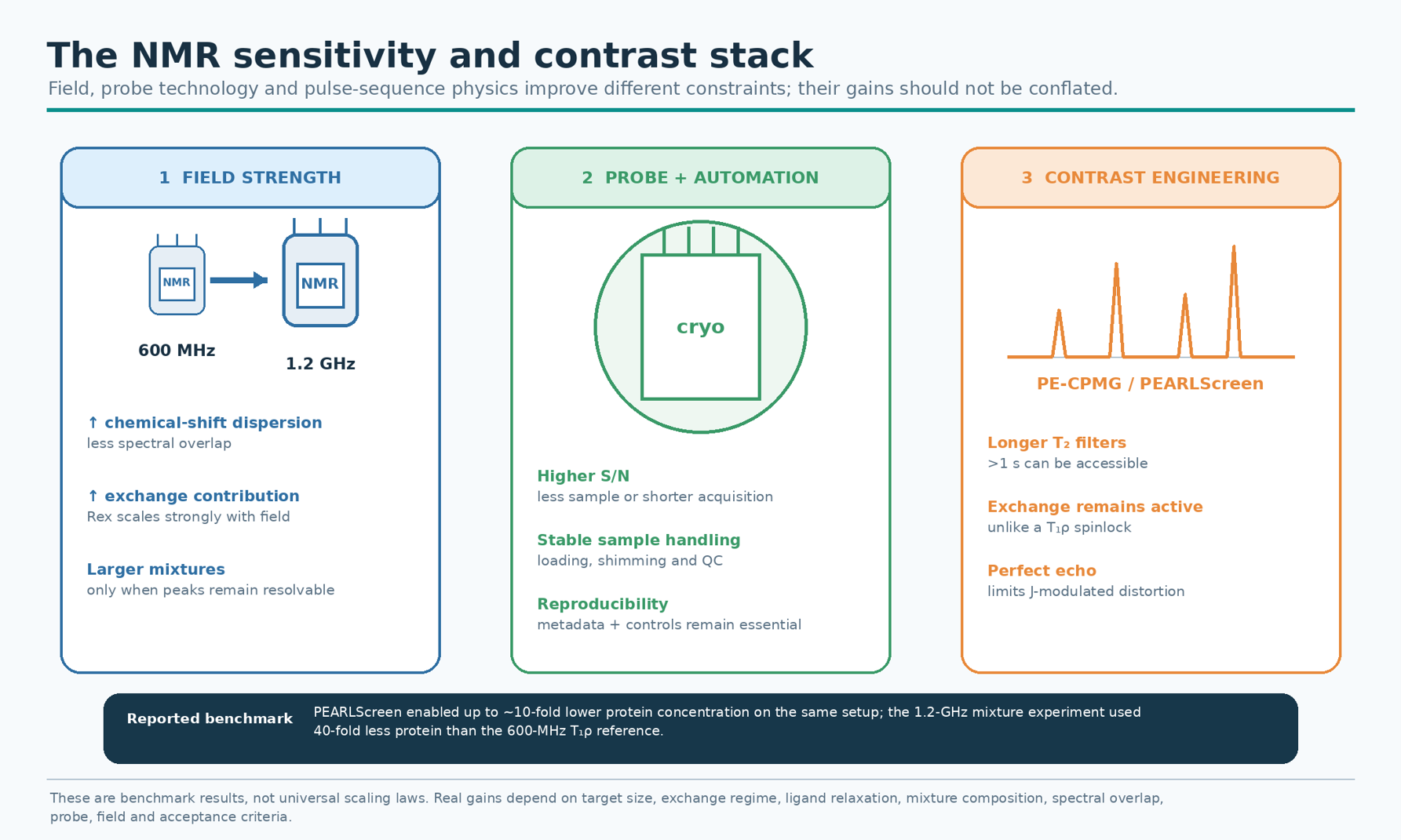
Figure 2. NMR sensitivity and contrast stack, showing how field strength, probe/automation performance, and pulse-sequence physics address different experimental constraints.
Ultra-High Field NMR And Contrast Engineering
Deployment reality — operational 1.2 GHz systems
At 28.2 T (1.2 GHz), ultra-high field provides major gains in spectral dispersion and practical sensitivity, alleviating spectral crowding and expanding the feasible experiment space — especially for protein-limited systems such as IDPs and membrane receptors. If installation counts are reported numerically, they should be tied to an explicit source and clearly separated into acquisition vs. commissioning vs. routine user operation.
PEARLScreen: Contrast as the right metric
Conventional ligand-observed screening experiments (T1ρ rotating-frame relaxation, waterLOGSY, and STD) suffer from inherent contrast limitations. T1ρ spinlock experiments suppress chemical shift differences between bound and free states, eliminating exchange-induced broadening that would otherwise enhance binding detection. Power deposition limits spinlock durations to 200 to 300 ms, constraining the relaxation window.
PEARLScreen addresses these limitations through perfect echo CPMG pulse trains. The perfect echo scheme prevents homonuclear J-coupling evolution — which would otherwise produce heavily distorted peak shapes with long inter-pulse delays — while allowing chemical exchange to contribute maximally to transverse relaxation.10 Relaxation times exceeding 1 second become accessible, versus the 200 to 300 ms ceiling for T1ρ experiments.
Benchmarking commercial spectrometers (80 to 1,200 MHz) showed a 10-fold protein reduction at standard fields and up to 40-fold at 1.2 GHz. In the reported 1.2 GHz benchmark, a 16-fragment mixture was screened with 40-fold lower protein consumption than the 600 MHz T₁ρ reference condition; this is a method-specific result rather than a universal scaling rule. For protein-limited FBDD targets, PEARLScreen with ultra-high field dramatically improves feasibility.
Read part 2 here.
References
- Hann MM, Leach AR, Harper G. Molecular complexity and its impact on the probability of finding leads for drug discovery. J Chem Inf Comput Sci 2001;41:856-864.
- Congreve M, Carr R, Murray C, Jhoti H. A 'rule of three' for fragment-based lead discovery? Drug Discov Today 2003;8:876-877.
- Hopkins AL, Groom CR, Alex A. Ligand efficiency: a useful metric for lead selection. Drug Discov Today 2004;9:430-431.
- Shuker SB, Hajduk PJ, Meadows RP, Fesik SW. Discovering high-affinity ligands for proteins: SAR by NMR. Science 1996;274:1531-1534.
- Feng BY, Simeonov A, Jadhav A, et al. A high-throughput screen for aggregation-based inhibition in a large compound library. J Med Chem 2007;50:2385-2390.
- Baell JB, Holloway GA. New substructure filters for removal of pan assay interference compounds (PAINS) from screening libraries. J Med Chem 2010;53:2719-2740.
- Dalvit C, Vulpetti A. Ligand-based fluorine NMR screening: principles and applications in drug discovery projects. J Med Chem 2019;62:2218-2244.
- Ohio State University. First 1.2 GHz NMR in U.S. installed at National Gateway Ultrahigh Field NMR Center. OSU News, December 19, 2023.
- Bruker Corporation. Swiss High-field NMR Facility acquires 1.2 GHz NMR at University of Zürich. Bruker Press Release, December 20, 2024.
- Lorz N, Czarniecki B, Loss S, Meier B, Gossert AD. Higher contrast in ¹H-observed NMR ligand screening with the PEARLScreen experiment. Angew Chem Int Ed 2025;64(17):e202423879.
- Binas O, de Jesus V, Landgraf T, et al. ¹⁹F NMR-based fragment screening for 14 different biologically active RNAs and 10 DNA and protein counter-screens. ChemBioChem 2021;22:423-433.
- de Castro GV, Ciulli A. Spy vs. spy: selecting the best reporter for ¹⁹F NMR competition experiments. Chem Commun 2019;55:1482-1485.
- Zhang Y, Zhu Y, Zhang Y, et al. ¹⁹F NMR chemical shift encoded peptide screening targeting the potassium channel Kv1.3. Chem Commun 2025;61:6162-6165.
- Gee CT, Arntson KE, Urick AK, et al. Protein-observed ¹⁹F-NMR for fragment screening, affinity quantification and druggability assessment. Nat Protoc 2016;11:1414-1427.
- Divakaran A, Kirberger SE, Pomerantz WCK. SAR by (protein-observed) ¹⁹F NMR. Acc Chem Res 2019;52:3407-3418.
- Fruh V, Zhou Y, Chen D, et al. Application of fragment-based drug discovery to membrane proteins: identification of ligands of the integral membrane enzyme DsbB. Chem Biol 2010;17:881-891.
- Medek A, Hajduk PJ, Mack J, Fesik SW. The use of differential chemical shifts for determining the binding site location and orientation of protein-bound ligands. J Am Chem Soc 2000;122:1241-1242.
- Dominguez C, Boelens R, Bonvin AM. HADDOCK: a protein-protein docking approach based on biochemical or biophysical information. J Am Chem Soc 2003;125:1731-1737.
- Kang C. Applications of in-cell NMR in structural biology and drug discovery. Int J Mol Sci 2019;20:139.
- Torres F, Bütikofer M, Stadler GR, et al. Ultrafast fragment screening using photo-hyperpolarized (CIDNP) NMR. J Am Chem Soc 2023;145:12066-12080.
About The Author
 Mahmoud K. Al-Ruweidi is a pharmaceutics specialist with expertise in rational drug design, discovery, and delivery. Trained as a biomedical engineer, his research spans formulation science and bioengineering approaches to medicine. He has worked across biochemistry, medical devices, and biomaterials, applying interdisciplinary methods to accelerate therapeutic innovation. Beyond the lab, he is an advocate for improving academic systems to better support young scientists and safeguard research integrity.