
Target Transcription Factor States, Not Pockets
By Mahmoud Khatib Al-Ruweidi

“Undruggable” is often less a property of a protein than a diagnosis of the method used to interrogate it. Transcription factors (TFs) expose broad protein–DNA and protein–protein interfaces, contain intrinsically disordered regions, and exchange partners as cellular context changes. A screen built around one purified, conformationally restricted domain can therefore be technically successful while missing the pharmacology that matters.1,2
Allostery offers a more useful framing because it changes the unit of intervention. A ligand need not occupy the DNA-recognition surface or the cofactor interface itself. It may instead redistribute the conformational ensemble that governs dimerization, cofactor recruitment, stability, or chromatin residence. The operative target is then not the TF sequence in isolation but a disease-relevant regulatory state linked to a measurable transcriptional consequence.3
This is not permission to describe every indirect intervention as allosteric. DNA-binding agents, epigenetic inhibitors, molecular glues, and degraders may all suppress TF-driven biology through different molecular events. The distinction is not semantic. Each mechanism creates different requirements for exposure, target engagement, resistance, and safety. A discovery program becomes more rigorous when its central claim is defined by the event it can demonstrate and falsify, rather than by the novelty of the label attached to it.
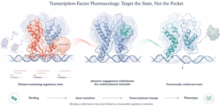
Figure 1. State-directed pharmacology of transcription factors. A disease-relevant transcription factor exists as a dynamic ensemble of regulatory states. Ligand engagement at a spatially distinct allosteric site can redistribute this ensemble, alter cofactor or complex stability, and redirect transcriptional output. The mechanistic claim therefore depends not only on binding but on demonstrating the predicted transition from target engagement to regulatory state control and phenotype.
The Target Is A Regulatory State
The discovery hypothesis should begin with the state that sustains disease. That means identifying the relevant isoform, oligomeric assembly, binding partner, post-translational modification, chromatin context, and subcellular compartment before screening begins. It also means deciding what the ligand is expected to do. Dimer dissociation, coactivator exclusion, mutant refolding, altered residence time, and destabilization of a transcriptionally competent complex are not interchangeable outcomes. They require different assays and imply different biomarkers.
A reporter gene alone is too distal to carry this argument. Transcription changes under stress, cytotoxicity, and broad chromatin disruption, often without specific control of the proposed TF state. A stronger target-state profile identifies a proximal pharmacodynamic readout, the magnitude and duration of modulation required for efficacy, the paralogs and normal tissues that define the safety boundary, and an experiment capable of disproving the mechanism. If the phenotype persists in a target-null cell or against an engagement-deficient allele, the state hypothesis has failed even when the reporter remains convincing.
Structure is essential here, but structure is evidence of possibility rather than proof of regulation. Allosteric pockets may be transient, ligand-induced, or absent from an isolated domain. Crystallography and cryogenic electron microscopy are therefore most informative when combined with methods that interrogate motion, including nuclear magnetic resonance, hydrogen–deuterium exchange mass spectrometry, molecular dynamics, and fragment-based mapping. Cryptic site prediction can locate openings that appear during protein motion, but only experiments can establish whether those openings persist, bind chemical matter, and communicate with function.4
Construct choice is consequently a mechanistic decision. A soluble domain may yield clean biophysical data while deleting the linker, disordered segment, partner, or modification that makes a pocket regulatory. Full-length protein and the disease-relevant complex should, where feasible, be studied alongside tractable domains rather than postponed until late validation. EN4 illustrates why this matters: it covalently engages Cys171 in an intrinsically disordered region of MYC and perturbs MYC transcriptional activity.5 The result is an important proof of principle, not evidence that disorder is generally easy to drug. Residue-level occupancy, proteome-wide selectivity, matched inactive analogs, and genetic resistance remain necessary to separate a functional covalent ligand from reactive chemistry.
The screening logic should follow the same progression. Binding or a change in local dynamics is the beginning of the case. The next question is whether that event shifts the specified regulatory state in a reconstituted complex, followed by whether the same transition occurs at achievable unbound concentrations in intact disease-relevant cells. Affinity against a convenient construct cannot compensate for flat potency in the native state. Nor can apparent selectivity be accepted without early paralog counter-screens, because a conserved cavity may exchange novelty for family-wide toxicity.
Evidence Must Follow The Mechanism
Transcription-factor programs are unusually capable of producing persuasive false positives because many cellular insults alter transcription. For that reason, the evidence cannot stop at binding, thermal stabilization, or loss of reporter signal. Direct interaction should be established by orthogonal biophysical methods and then demonstrated in intact cells using a cellular thermal shift assay, bioluminescence resonance energy transfer, or a competitive chemoproteomic probe.6,7 These measurements answer whether the compound reaches and engages the TF. They do not yet establish what engagement does.
Mechanism requires the predicted state transition. A dimer-disrupting ligand should alter partner stoichiometry, a conformational stabilizer should restore the relevant structural state, and a chromatin-residence modulator should change occupancy at prespecified loci. Chromatin immunoprecipitation sequencing or CUT&RUN can then connect that proximal event to a defined transcriptional program.8 Target depletion, an engagement-deficient mutant, or rescue with a compound-insensitive allele closes the causal chain between binding, state control, transcription, and phenotype. This order matters because it separates mechanism from consequence rather than inferring one from the other.
Concentration and time must also remain visible across the chain. Biochemical affinity, cellular occupancy, state transition potency, transcriptional response, and phenotype are best compared on the same unbound concentration scale. A large disconnect may reveal poor permeability, intracellular sequestration, active efflux, nonspecific binding, or an off-target stress response. Washout experiments add a temporal dimension: when transient occupancy produces durable chromatin or transcriptional effects, they can distinguish continuous target coverage from a pulse that resets the regulatory state.
Mechanistic nomenclature should reflect this evidence. An allosteric modulator binds the TF at a functionally coupled site. An orthosteric inhibitor blocks the operative interface directly. A molecular glue creates or stabilizes a new interface, while a degrader changes protein abundance through induced proximity. DNA-binding agents and epigenetic inhibitors act at still different levels of the regulatory system. These modalities may converge phenotypically, but they are not pharmacologically equivalent. Collapsing them into a single category conceals the exposure, kinetic, resistance, and safety questions that determine whether a program can translate.

What Clinical Precedents Actually Teach
Hypoxia-inducible factor 2α (HIF-2α) provides the clearest clinical validation of state-directed TF pharmacology. Small molecules bind a cavity in the PAS-B domain and disrupt heterodimerization with ARNT through an allosteric mechanism.9 Preclinical studies connected this state transition to tumor dependency, and belzutifan subsequently produced clinical responses in von Hippel–Lindau disease-associated tumors.10,11 Yet the same program also revealed the vulnerability of pocket-dependent selectivity: the EPAS1 G323E mutation impairs inhibitor binding and can drive acquired resistance.12 The achievement was therefore not simply the discovery of a hidden cavity. It was the connection of cavity occupancy to complex dissociation, genotype-defined dependence, pharmacodynamic measurement, and a clinically meaningful therapeutic window.
Transcriptional enhanced associate domain (TEAD) pharmacology offers a related but distinct model. Structural studies identified a central lipid-binding cavity, while work on TEAD autopalmitoylation established that this pocket is functionally coupled to Hippo-pathway output.13,14 The oral inhibitor VT3989 has now provided early clinical proof of concept in mesothelioma and other solid tumors.15 The broader lesson is that endogenous regulatory pockets, including lipid-filled cavities, deserve systematic screening. The qualification is equally important: TEAD isoforms contribute to normal tissue homeostasis, so pocket conservation, isoform selectivity, and the tolerability of sustained pathway suppression cannot be treated as downstream questions.
Mutant p53 reverses the usual direction of the intervention. The Y220C substitution creates a crevice but also destabilizes the DNA-binding domain. Rezatapopt occupies this mutant-specific site, restores a wild-type-like conformation and transcriptional function, and has produced objective responses in a Phase 1 study.16,17 Here the desired state is rescued rather than inhibited. The allele restriction of the drug, together with the association between response and KRAS wild-type tumors, shows why a structural biomarker is necessary but not sufficient. Network context remains part of the pharmacology.
These precedents should not be compressed into a generic claim that TFs are now druggable. They represent different value propositions: dissociating a disease-sustaining complex, perturbing an endogenous regulatory pocket, and rescuing a mutant conformation. Each demands its own assay architecture and patient selection logic. BCL6 sharpens the point. Ligands of its BTB domain may inhibit corepressor binding, whereas BI-3802 induces BCL6 polymerization followed by degradation.18,19 Similar chemical matter can therefore generate fundamentally different events. The mechanism is whatever the data establish, not whatever the program was designed to find.
Translation Begins Before Lead Optimization
A technically interesting binder is not yet a development hypothesis. Before extensive lead optimization, the relevant state should be demonstrated in human disease material, its modulation should produce a therapeutic window, and the proximal biomarker should be measurable in preclinical species and, eventually, patients. The required tissue exposure must also be compatible with the compound’s physicochemical properties and safety margin. Nuclear localization is not a unique barrier for a permeable small molecule, but high nonspecific binding, active efflux, and lysosomal trapping can make nominal cellular potency look more persuasive than the achievable free concentration.
Translation is the point at which the mechanism must survive changes of scale. A state transition observed in a purified system must remain detectable in intact cells and human disease material; the phenotype must depend on target engagement; and the effective unbound concentration must remain compatible with plausible tissue exposure. Binding, state change, chromatin occupancy, transcriptional response, and phenotype do not need to produce identical numerical values, but their relationship must be explainable. An unexplained separation between them is not experimental noise to be averaged away. It is often the first indication that the proposed mechanism is incomplete.
Resistance belongs inside this assessment rather than after it. Allosteric selectivity often depends on a small number of pocket residues, creating an efficient route to escape.20 Saturation mutagenesis or base-editing scans can expose vulnerable positions before candidate nomination, while resistant population sequencing can distinguish pocket escape from partner switching, paralog compensation, lineage plasticity, altered chromatin accessibility, or loss of the biomarker-defined dependency. These bypass states may preserve transcription without changing the pocket at all.
The same information should shape combination strategy. Two agents do not form a rational combination merely because both reduce growth. A defensible pairing blocks a measured escape route, preserves the therapeutic window, and carries biomarkers that distinguish loss of target control from pathway bypass. Second-generation chemistry should also begin before resistance becomes a clinical observation. Chemically distinct scaffolds, mapped substitutions around resistance-prone residues, and assays that separate loss of exposure from loss of state control turn resistance from a retrospective explanation into a discovery variable.
A Better Unit Of Discovery
The most consequential shift in TF drug discovery is therefore conceptual and operational: treat the regulatory state as the target, then construct the chemistry, assays, biomarkers, and resistance studies around that state. This approach will not make every TF tractable, nor should it. Its value is more disciplined. It replaces the binary of druggable versus undruggable with testable questions about conformational coupling, disease dependence, exposure, and causality. That is a stronger basis for deciding which difficult targets deserve a program and which merely possess an interesting pocket.
References
- Bushweller, J.H. Targeting transcription factors in cancer — from undruggable to reality. Nat. Rev. Cancer 19, 611–624 (2019). https://doi.org/10.1038/s41568-019-0196-7.
- Henley, M.J. & Koehler, A.N. Advances in targeting ‘undruggable’ transcription factors with small molecules. Nat. Rev. Drug Discov. 20, 669–688 (2021). https://doi.org/10.1038/s41573-021-00199-0.
- Motlagh, H.N., Wrabl, J.O., Li, J. & Hilser, V.J. The ensemble nature of allostery. Nature 508, 331–339 (2014). https://doi.org/10.1038/nature13001.
- Cimermancic, P. et al. CryptoSite: Expanding the druggable proteome by characterization and prediction of cryptic binding sites. J. Mol. Biol. 428, 709–719 (2016). https://doi.org/10.1016/j.jmb.2016.01.029.
- Boike, L. et al. Discovery of a functional covalent ligand targeting an intrinsically disordered cysteine within MYC. Cell Chem. Biol. 28, 4–13.e17 (2021). https://doi.org/10.1016/j.chembiol.2020.09.001.
- Martinez Molina, D. et al. Monitoring drug target engagement in cells and tissues using the cellular thermal shift assay. Science 341, 84–87 (2013). https://doi.org/10.1126/science.1233606.
- Robers, M.B. et al. Target engagement and drug residence time can be observed in living cells with BRET. Nat. Commun. 6, 10091 (2015). https://doi.org/10.1038/ncomms10091.
- Skene, P.J. & Henikoff, S. An efficient targeted nuclease strategy for high-resolution mapping of DNA binding sites. eLife 6, e21856 (2017). https://doi.org/10.7554/eLife.21856.
- Scheuermann, T.H. et al. Allosteric inhibition of hypoxia-inducible factor 2 with small molecules. Nat. Chem. Biol. 9, 271–276 (2013). https://doi.org/10.1038/nchembio.1185.
- Chen, W. et al. Targeting renal cell carcinoma with a HIF-2 antagonist. Nature 539, 112–117 (2016). https://doi.org/10.1038/nature19796.
- Jonasch, E. et al. Belzutifan for renal cell carcinoma in von Hippel–Lindau disease. N. Engl. J. Med. 385, 2036–2046 (2021). https://doi.org/10.1056/NEJMoa2103425.
- Courtney, K.D. et al. HIF-2 complex dissociation, target inhibition, and acquired resistance with PT2385 in patients with clear cell renal cell carcinoma. Clin. Cancer Res. 26, 793–803 (2020). https://doi.org/10.1158/1078-0432.CCR-19-1459.
- Pobbati, A.V. et al. Targeting the central pocket in human transcription factor TEAD as a potential cancer therapeutic strategy. Structure 23, 2076–2086 (2015). https://doi.org/10.1016/j.str.2015.09.009.
- Chan, P. et al. Autopalmitoylation of TEAD proteins regulates transcriptional output of the Hippo pathway. Nat. Chem. Biol. 12, 282–289 (2016). https://doi.org/10.1038/nchembio.2036.
- Yap, T.A. et al. YAP/TEAD inhibitor VT3989 in solid tumors: A Phase 1/2 trial. Nat. Med. 31, 4281–4290 (2025). https://doi.org/10.1038/s41591-025-04029-3.
- Puzio-Kuter, A.M. et al. Restoration of the tumor suppressor function of Y220C-mutant p53 by rezatapopt, a small-molecule reactivator. Cancer Discov. 15, 1159–1179 (2025). https://doi.org/10.1158/2159-8290.CD-24-1421.
- Dumbrava, E.E. et al. Phase 1 study of rezatapopt, a p53 reactivator, in TP53 Y220C-mutated tumors. N. Engl. J. Med. 394, 872–883 (2026). https://doi.org/10.1056/NEJMoa2508820.
- Cerchietti, L.C. et al. A small-molecule inhibitor of BCL6 kills DLBCL cells in vitro and in vivo. Cancer Cell 17, 400–411 (2010). https://doi.org/10.1016/j.ccr.2009.12.050.
- Słabicki, M. et al. Small-molecule-induced polymerization triggers degradation of BCL6. Nature 588, 164–168 (2020). https://doi.org/10.1038/s41586-020-2925-1.
- Lu, S. et al. Emergence of allosteric drug-resistance mutations: New challenges for allosteric drug discovery. Drug Discov. Today 25, 177–184 (2020). https://doi.org/10.1016/j.drudis.2019.10.006.
About The Author
 Mahmoud K. Al-Ruweidi is a pharmaceutics specialist with expertise in rational drug design, discovery, and delivery. Trained as a biomedical engineer, his research spans formulation science and bioengineering approaches to medicine. He has worked across biochemistry, medical devices, and biomaterials, applying interdisciplinary methods to accelerate therapeutic innovation. Beyond the lab, he is an advocate for improving academic systems to better support young scientists and safeguard research integrity.