
Measuring Cellular Water Content As A New Tool For Organoid Characterization
By Georgios Katsikis, Ph.D., CTO, Drosera Biotechnologies, and Teemu P. Miettinen, Ph.D., Research Scientist, Koch Institute for Integrative Cancer Research, Massachusetts Institute of Technology

Human-based organoids and tumor spheroids have emerged as key disease models with higher physiological relevance over two-dimensional cell cultures.1 The increased interest in organoids and other 3D model systems was also highlighted by the FDA’s recent announcement to phase out animal testing for drug development.2 As these models mature, the challenge for their broader adoption involves generating and holistically characterizing their biological state. Current characterization workflows assess organoid morphology, molecular composition, and function.3 Yet one of the most fundamental determinants of cell physiology, the cellular water content, which governs macromolecular concentrations and reaction rates and is involved in many disease phenotypes,4–6 has remained unstudied in living organoids and other similar 3D model systems.
A collaboration between MIT, KAIST, and Dana Farber Cancer Institute created a platform technology (Fig. 1) that, for the first time, quantifies water content in single organoids. We leveraged the precision of our platform (~0.5% error in water content measurement) to resolve heterogeneity in water content across individual patient-derived glioblastoma spheroids, as well as characterize acute water content changes in response to drug treatment — findings that were previously inaccessible.
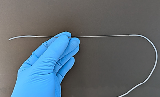
Fig. 1. Photograph of steel tube used for measuring the water content of living organoids using a steel tube
Water is the basis of life as the medium in which biochemistry operates. Within any given cell, concentrations of macromolecules and their interaction rates are inversely correlated with water content, whereas molecular transport rates via diffusion are directly correlated with water content (Fig. 2). This relationship creates evolutionary pressure for cells to regulate their intracellular water content to optimize cellular functions.7 While this fundamental importance of cellular water content is well recognized and often discussed as molecular crowding,8 surprisingly little is known about how water content is regulated and how various perturbations, such as pharmaceutical interventions, influence cellular water content. This is especially true for physiologically relevant model systems, such as tumor spheroids or organoids, which have been shown to be sensitive to changes in intracellular crowding and water content.9
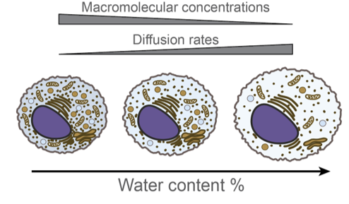
Fig. 2. Schematic of intracellular water content and the molecular consequences of changed water content.
This knowledge gap in the regulatory role of water content is attributed to the lack of approaches that can quantitatively measure intracellular water. We and others have developed technologies that can measure water content in isolated cells or 2D tissue layers,10–13 but such technologies were not designed to study larger 3D samples.
Our technology works by flowing the sample, such as an organoid, inside a steel tube that is vibrating at its resonant frequency (Fig. 3). When the sample flows through the tube, it displaces water and makes the tube heavier. The tube behaves similarly to a vibrating guitar string; the heavier the string is, the lower frequency (more base sound) signal it produces. In our tube, this signal is not audible (resonant frequencies are an order of magnitude higher than audible range), but we electronically capture a change in the resonant frequency when the sample flows through.
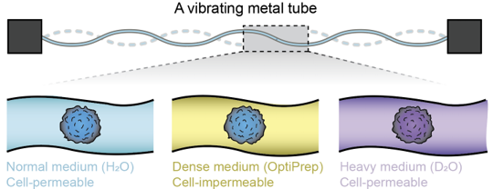
Fig. 3. Schematic of the measurement concept, where a vibrating steel tube is used to sense the buoyant mass of the sample (e.g., an organoid) in different fluids to determine the sample’s water content.
The key function is that a given sample affects the resonant frequency proportionally to the change in mass on the system, and this enables us to determine the buoyant mass of the sample. Following this, we change the media surrounding the sample and repeat the measurement (Fig. 2). By sequentially measuring a given sample in different media, some of which are cell permeable and some of which are not, we can back calculate its absolute and relative water content simultaneously with other biophysical properties, such as sample volume, dry mass, and density.

The measurement technology is simple. It is not based on a cleanroom-fabricated microfluidic device but rather a repurposed robust steel tube. We have proved that the measurement works in sub-mm size ranges, but the system is amenable to customization at different sample sizes. This scalability to different size ranges and the broadly applicable water content measurement principle enable our method to be used, in principle, with any 3D model system that can be maintained in suspension, including (small) whole organism drug discovery models, such as C. elegans. The measurements consist of single scalar outputs, which are immune to user-specific interpretations, unlike many imaging-based approaches. Notably, among these outputs are sample volume and mass, which can be detected with far greater precision (e.g., <1% error) than with conventional microscopy-based approaches. Because changes in size and mass are fundamental indicators of growth and drug response, these measurements provide a direct quantitative means of assessing treatment efficacy. In addition, we experimentally demonstrated that measurements do not compromise the viability of tumor spheroids. The measurements are flow through with commercially available media components, and the measurements can be integrated with robotic pipette systems for downstream sample collection and integration with different characterization modalities, such as drug testing, morphological imaging, and omics analyses.
Ultimately, the probability of successful drug discovery not only depends on the number of candidates screened but also on the quality of the experimental methods used to identify the most promising candidates.14 We see our technology as a strong fit for the new approach methodologies that the FDA is now actively encouraging. As demonstrated by the FDA's ISTAND program, this is a rapidly expanding space spanning wet-bench platforms (organoids and organ-on-chip systems), imaging, transcriptomics, and proteomics, alongside in silico biosimulation.15 Spheroids and organoids — including vascularized formats16 — are prime examples of the model systems our platform can measure, providing quantitative and orthogonal insights into existing techniques.
The results were published on April 3, 2026, in Science Advances.
References
- S. Gunti, A. T. K. Hoke, K. P. Vu, N. R. London, Organoid and Spheroid Tumor Models: Techniques and Applications. Cancers (Basel). 13, 1–18 (2021).
- FDA Announces Plan to Phase Out Animal Testing Requirement for Monoclonal Antibodies and Other Drugs | FDA. https://www.fda.gov/news-events/press-announcements/fda-announces-plan-phase-out-animal-testing-requirement-monoclonal-antibodies-and-other-drugs.
- Y. Gu, W. Zhang, X. Wu, Y. Zhang, K. Xu, J. Su, Organoid assessment technologies. Clin. Transl. Med. 13 (2023).
- P. G. Gallagher, Disorders of erythrocyte hydration. Blood 130, 2699–2708 (2017).
- A. S. Verkman, Aquaporins in clinical medicine. Annu. Rev. Med. 63, 303–316 (2012).
- M. D. Sorani, Z. Zado, E. Hurowitz, D. Yan, K. M. Giacomini, G. T. Manley, Novel variants in human Aquaporin-4 reduce cellular water permeability. Hum. Mol. Genet. 17, 2379–2389 (2008).
- J. H. Huang, J. E. Ferrell, How does cytoplasmic crowding affect reaction rates? Mol. Cell 86, 9–23 (2026).
- C. Alfano, Y. Fichou, K. Huber, M. Weiss, E. Spruijt, S. Ebbinghaus, G. De Luca, M. A. Morando, V. Vetri, P. A. Temussi, A. Pastore, Molecular Crowding: The History and Development of a Scientific Paradigm. Chem. Rev. 124, 3186–3219 (2024).
- Y. Li, M. Chen, J. Hu, R. Sheng, Q. Lin, X. He, M. Guo, Volumetric Compression Induces Intracellular Crowding to Control Intestinal Organoid Growth via Wnt/β-Catenin Signaling. Cell Stem Cell 28, 63-78.e7 (2021).
- F. Feijó Delgado, N. Cermak, V. C. Hecht, S. Son, Y. Li, S. M. Knudsen, S. Olcum, J. M. Higgins, J. Chen, W. H. Grover, S. R. Manalis, Intracellular Water Exchange for Measuring the Dry Mass, Water Mass and Changes in Chemical Composition of Living Cells. PLoS One 8 (2013).
- Y. Wu, V. K. Kouadio, T. R. Usherwood, J. Li, M. Bisher, R. Aurora, A. Z. Lam, A. R. Lam, A. K. R. Lytton-Jean, S. R. Manalis, T. P. Miettinen, Diverse biophysical and molecular mechanisms drive phytoplankton sinking in response to starvation. PLoS Biol. 23, e3003508 (2025).
- S. Oh, C. H. Lee, W. Yang, A. Li, A. Mukherjee, M. Basan, C. Ran, W. Yin, C. J. Tabin, D. Fu, X. S. Xie, M. W. Kirschner, Protein and lipid mass concentration measurement in tissues by stimulated Raman scattering microscopy. Proc. Natl. Acad. Sci. U. S. A. 119, e2117938119 (2022).
- T. P. Miettinen, A. L. Gomez, Y. Wu, W. Wu, T. R. Usherwood, Y. Hwang, B. R. K. Roller, M. F. Polz, S. R. Manalis, Cell size, density, and nutrient dependency of unicellular algal gravitational sinking velocities. Sci. Adv. 10, 8356 (2024).
- J. W. Scannell, J. Bosley, J. A. Hickman, G. R. Dawson, H. Truebel, G. S. Ferreira, D. Richards, J. M. Treherne, Predictive validity in drug discovery: what it is, why it matters and how to improve it. Nat. Rev. Drug Discov. 21, 915–931 (2022).
- C. Sheridan, AI-powered methods shake up animal testing. Nat. Biotechnol. 44, 1067–1070 (2026).
- A. de Janon, P. Kaza, K. Yoshida, S. J. Lee, D. DeRyckere, D. K. Graham, S. Takayama, Free-floating long-term vascularized mesenchymal organoids. iScience 29 (2026).
About The Authors:
 Georgios ‘Yorgos’ Katsikis is chief technology officer at Drosera Biotechnologies, a Cambridge, MA-based biotechnology company. He is an inventor and developer of automation technologies for the characterization and processing of biological systems, ranging from viral vectors to cells and 3D cell models. He was previously VP of engineering at Anthology Biotechnologies. He earned his Ph.D. in mechanical engineering from Stanford University. He can be reached via LinkedIn or at katsikis.g@gmail.com.
 Teemu P. Miettinen, Ph.D., is a research scientist at the Koch Institute for Integrative Cancer Research at the Massachusetts Institute of Technology. His research seeks to understand how cell size, growth, and composition are regulated and interconnected across a variety of model systems. He also works as a part of the laboratory of Professor Scott Manalis, where they jointly develop novel biophysical measurements for biomedical applications. He has authored several dozen research papers and two patents. He can be reached at teemu@mit.edu.