
Beyond Apoptosis: Context Is The Target
By Mahmoud Khatib Al-Ruweidi

Apoptosis gave drug discovery a tractable language for killing diseased cells: identify a survival dependency, release the execution machinery, and measure whether the cell dies. The success of BCL-2 inhibition showed what becomes possible when mechanism, biomarker, and clinical context align. It also exposed the limit of treating cell death as a single pathway. Cells that evade apoptosis do not necessarily evade death. They may remain vulnerable to ferroptosis, pyroptosis, necroptosis, or another regulated cell death program.
The expanding nomenclature is scientifically useful, but it can mislead discovery teams into treating each death modality as a self-contained target class. It is not. A pathway name describes molecular execution; it does not specify which cell should die, when death should occur, what neighboring cells will experience, or whether the tissue-level effect will be therapeutic. The useful discovery unit is therefore not ferroptosis or pyroptosis in isolation. It is a causal system linking exposure, cell state, execution mechanism, immune consequence, and tissue response.1,2
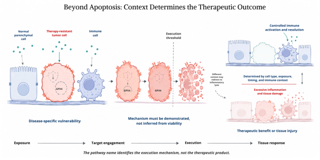
Figure 1. Context determines the therapeutic outcome of regulated cell death. A disease-associated cellular state may create selective vulnerability to a defined death mechanism, but pathway engagement alone does not establish therapeutic value. The outcome depends on exposure in the relevant compartment, execution within the intended cell population, preservation of normal and immune cell function, and the resulting tissue response. The same molecular mechanism may therefore produce selective disease cell elimination or clinically limiting tissue injury.
The Pathway Name Is Not the Product
Regulated cell death is directionally ambivalent. In an apoptosis-resistant tumor, inducing lipid peroxidation may eliminate a persister population. In the kidney, brain, or heart, the same chemistry can destroy irreplaceable parenchymal cells. Gasdermin pore formation may convert tumor cell death into an inflammatory signal, yet the same execution step can amplify septic or sterile inflammation. RIPK1 inhibition may protect tissue in one setting while weakening host defense circuitry in another.
This is why the indication alone cannot define the pharmacology. Before screening compounds, a program should specify the target compartment and the intended causal chain: which cell state carries the vulnerability; which molecular step must be engaged; whether the desired effect is cell elimination, temporary suppression of lysis, or interruption of inflammatory propagation; and which neighboring cells must remain pharmacologically untouched. A ferroptosis program in cancer, for example, should distinguish malignant cell lipid peroxide liability from the vulnerability of infiltrating lymphocytes and normal tissue. “Induce ferroptosis in the tumor” is not yet a product concept.
The same discipline applies to newly named modalities. Cuproptosis established that copper can kill cells through a mitochondrial, lipoylated-protein-dependent mechanism, while PANoptosis describes coordinated engagement of pyroptotic, apoptotic, and necroptotic machinery.3,4 These advances expand the mechanistic map. They do not remove the need to demonstrate that the mapped mechanism controls the disease-relevant outcome.
Prove Mechanism Before Optimizing Potency
A viability signal does not identify a death mechanism. ATP depletion, membrane-impermeant dyes, Annexin V staining, or a single cleaved protein can report parts of a trajectory, but each can also arise downstream of several lethal processes. The Nomenclature Committee on Cell Death therefore favors mechanistic definitions, and assay guidance increasingly emphasizes convergent evidence rather than marker-based labeling.1,5
Discovery teams should build that convergence into the primary cascade. Start with time-resolved evidence that cells actually die, then connect death to the proposed execution machinery using perturbations that answer different questions. Chemical rescue tests pharmacological reversibility. Genetic perturbation tests dependency. Biochemical or imaging assays establish target engagement and the predicted downstream event. Long-term regrowth or clonogenic recovery determines whether an apparent rescue prevented death or merely delayed it.
The exact package must fit the mechanism. A ferroptosis claim should combine lethal lipid peroxidation with iron dependence and rescue by a mechanistically appropriate radical trapping agent; it should also use genetic perturbations matched to the initiating stimulus. That last qualification matters. An integration of 24 ferroptosis screens found that genetic dependencies vary substantially with cellular context and with the way ferroptosis is initiated. ACSL4, for instance, was more important after direct GPX4 inhibition than after cystine deprivation.6 A universal “ferroptosis signature” can therefore conceal the variable that actually governs response.
For pyroptosis, gasdermin cleavage alone is insufficient because processing and pore formation are separable. Disulfiram can permit GSDMD and interleukin-1β processing while preventing GSDMD pore formation, cytokine release, and lysis.7 Gasdermin-targeting nanobodies reinforce the same principle: downstream execution can be pharmacologically modulated without erasing every upstream inflammatory event.8 A necroptosis claim likewise requires more than RIPK1 or MLKL phosphorylation. It should establish RIPK3–MLKL dependency, membrane execution, and rescue under conditions that do not simply divert cells into apoptosis or another form of lytic death.9
Morphology remains useful, particularly when it reveals membrane rupture, organelle swelling, or ferroptosis-associated mitochondrial changes. It should support a mechanistic case, not substitute for one.
Kinetics Exposes What Endpoint Assays Hide
Two compounds can produce the same 48-hour viability value through very different pharmacology. One may kill nearly all exposed cells slowly. Another may kill a susceptible fraction within hours and leave a stable persister population. A third may suppress proliferation without causing death. Endpoint assays collapse onset, rate, and lethal fraction into one number.
Time-lapse measurements separate those variables and reveal when rescue occurs, whether death propagates between cells, and whether a blocked pathway reappears through another execution route. Scalable analysis of population death kinetics has shown that potent lethal compounds produce widely divergent temporal profiles even within the same cell system.10 That information should shape concentration selection, combination timing, and biomarker sampling. If target engagement peaks after the decisive execution event, a pharmacodynamic biopsy may be analytically precise and biologically uninformative.
Kinetic analysis also changes how combinations should be screened. A favorable endpoint interaction may reflect faster killing rather than a larger lethal fraction; the distinction affects durability. Conversely, an early rescue may disappear at later time points because the cell switches pathways. Combination matrices should therefore include temporal response surfaces and washout or regrowth phases, not a single synergy score.

Treat Pathway Switching As A Design Variable
Cell death pathways share regulatory nodes. Caspase-8 can restrain necroptosis while enabling apoptotic execution. Caspase-3 can produce apoptotic morphology in one cell but activate GSDME-dependent pyroptosis in another. Membrane repair machinery can delay lysis after gasdermin or MLKL engagement. Blocking one route may consequently protect the cell, change the kinetics, or redirect death into a more inflammatory form.
Discovery programs should test these alternatives deliberately. Run key perturbations in genetically defined backgrounds, profile at least the principal competing execution pathways, and measure the extracellular consequence rather than assuming that more lysis is therapeutically better. In oncology, this means assessing tumor cell killing alongside cytokine and damage-associated molecular pattern release, dendritic cell activation, and lymphocyte fitness. In inflammatory disease, it means determining whether a pore blocker reduces pathological lysis while preserving antimicrobial functions that occur upstream or through parallel pathways.
This systems view also sharpens synthetic lethality claims. Drug-tolerant persister cells can acquire a dependency on GPX4, creating a real opportunity to bypass resistance.11 But the actionable biomarker is not “persister” as a broad label. It is the lipid and antioxidant state that creates GPX4 dependence, together with the exposure window in which that state persists. The combination should be scheduled around that induced vulnerability rather than administered merely because two agents kill more cells together.
Translate A Causal Chain, Not A Marker
Clinical translation fails when biomarkers occupy only one end of the mechanism. Target engagement without evidence of execution cannot show that the intended death program occurred. A circulating damage marker without target compartment resolution cannot distinguish tumor killing from normal tissue injury. Conversely, a pathway marker may be biologically real yet clinically irrelevant if the tissue repairs the lesion or the immune microenvironment reverses the benefit.
The biomarker strategy should follow the causal chain: exposure in the relevant compartment, engagement of the proposed control node, activation or inhibition of the execution step, change in the intended cell population, and a tissue-level consequence linked to the clinical objective. The chain will rarely rely on one analyte. It may require drug concentration; a proximal target engagement assay; an execution marker, such as oxidized phospholipid species or membrane-localized MLKL; and a spatial or immune readout. The purpose is not maximal assay density. It is to identify where the pharmacology succeeds or fails.
This framework changes lead optimization as well. Potency against a death regulator is only one attribute. Distribution, residence time, cell-selective uptake, and reversibility may determine whether the same molecule becomes an antitumor agent or a tissue toxicant. Ferroptosis illustrates the problem clearly: genetic loss of GPX4 can cause acute organ injury, while GPX4 dependence can also create a vulnerability in therapy-resistant cancer cells. 11,12 Medicinal chemistry and delivery strategy must therefore optimize spatial and temporal selectivity together with biochemical potency. A leading translational road map reaches the same conclusion: pharmacology must resolve context before it can exploit mechanism.13
Regulated cell death should move drug discovery beyond an apoptosis-centric model, but not into a catalogue of fashionable pathway names. The decisive questions are more demanding: which cells are being pushed across which threshold, under what exposure, through which execution machinery, with what consequence for the surrounding tissue? Programs that answer those questions early will discard attractive but noncausal phenotypes, design more informative combinations, and build biomarkers that can survive the transition from a culture dish to a patient.
References
1. Galluzzi L, Vitale I, Aaronson SA, et al. Molecular mechanisms of cell death: recommendations of the Nomenclature Committee on Cell Death 2018. Cell Death Differ. 2018;25(3):486–541. doi:10.1038/s41418-017-0012-4.
2. Yuan J, Ofengeim D. A guide to cell death pathways. Nat Rev Mol Cell Biol. 2024;25(5):379–395. doi:10.1038/s41580-023-00689-6.
3. Tsvetkov P, Coy S, Petrova B, et al. Copper induces cell death by targeting lipoylated TCA cycle proteins. Science. 2022;375(6586):1254–1261. doi:10.1126/science.abf0529.
4. Lee S, Karki R, Wang Y, Nguyen LN, Kalathur RC, Kanneganti TD. AIM2 forms a complex with pyrin and ZBP1 to drive PANoptosis and host defence. Nature. 2021;597(7876):415–419. doi:10.1038/s41586-021-03875-8.
5. Hu X, Li Z, Lin R, et al. Guidelines for regulated cell death assays: a systematic summary, a categorical comparison, a prospective. Front Cell Dev Biol. 2021;9:634690. doi:10.3389/fcell.2021.634690.
6. Magtanong L, Mueller GD, Williams KJ, et al. Context-dependent regulation of ferroptosis sensitivity. Cell Chem Biol. 2022;29(9):1409–1418.e6. doi:10.1016/j.chembiol.2022.06.004.
7. Hu JJ, Liu X, Xia S, et al. FDA-approved disulfiram inhibits pyroptosis by blocking gasdermin D pore formation. Nat Immunol. 2020;21(7):736–745. doi:10.1038/s41590-020-0669-6.
8. Kopp A, Hagelueken G, Jamitzky I, et al. Pyroptosis inhibiting nanobodies block gasdermin D pore formation. Nat Commun. 2023;14(1):7923. doi:10.1038/s41467-023-43707-z.
9. Sun L, Wang H, Wang Z, et al. Mixed lineage kinase domain-like protein mediates necrosis signaling downstream of RIP3 kinase. Cell. 2012;148(1–2):213–227. doi:10.1016/j.cell.2011.11.031.
10. Forcina GC, Conlon M, Wells A, Cao JY, Dixon SJ. Systematic quantification of population cell death kinetics in mammalian cells. Cell Syst. 2017;4(6):600–610.e6. doi:10.1016/j.cels.2017.05.002.
11. Hangauer MJ, Viswanathan VS, Ryan MJ, et al. Drug-tolerant persister cancer cells are vulnerable to GPX4 inhibition. Nature. 2017;551(7679):247–250. doi:10.1038/nature24297.
12. Friedmann Angeli JP, Schneider M, Proneth B, et al. Inactivation of the ferroptosis regulator GPX4 triggers acute renal failure in mice. Nat Cell Biol. 2014;16(12):1180–1191. doi:10.1038/ncb3064.
13. Hadian K, Stockwell BR. A roadmap to creating ferroptosis-based medicines. Nat Chem Biol. 2021;17(11):1113–1116. doi:10.1038/s41589-021-00853-z.
About The Author
 Mahmoud K. Al-Ruweidi is a pharmaceutics specialist with expertise in rational drug design, discovery, and delivery. Trained as a biomedical engineer, his research spans formulation science and bioengineering approaches to medicine. He has worked across biochemistry, medical devices, and biomaterials, applying interdisciplinary methods to accelerate therapeutic innovation. Beyond the lab, he is an advocate for improving academic systems to better support young scientists and safeguard research integrity.