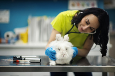
In Vivo And Histological Analysis Of Focal Chorioretinal Defects In Dutch Belted Rabbits
By Simone Iwabe, Stephanie Michelle Shrader, Stefan Linehan, Jeffrey Burdick, and Gustavo David Aguirre

Baseline ophthalmic examinations, including slit lamp and indirect ophthalmoscope assessments, are essential for evaluating ocular health across all species involved in ocular toxicity studies. These exams are crucial for identifying spontaneous ocular lesions, which have been reported during prescreening in various species, such as mice, rats, dogs, and nonhuman primates. These lesions can involve the cornea, iris, lens, vitreous, and retina. In addition to these species, laboratory rabbits—particularly the Dutch Belted and New Zealand White breeds—are also commonly used in ocular toxicology studies. However, there is limited data on spontaneous ocular lesions in rabbits, with most reports focusing on the cornea, iris, lens, and vitreous.The goal of our study is to explore the in vivo retinal microanatomy in Dutch Belted
laboratory rabbits, specifically examining findings observed during pretest ophthalmic examinations, to better understand potential spontaneous ocular lesions in this species.
Get unlimited access to:
Enter your credentials below to log in. Not yet a member of Drug Discovery Online? Subscribe today.
